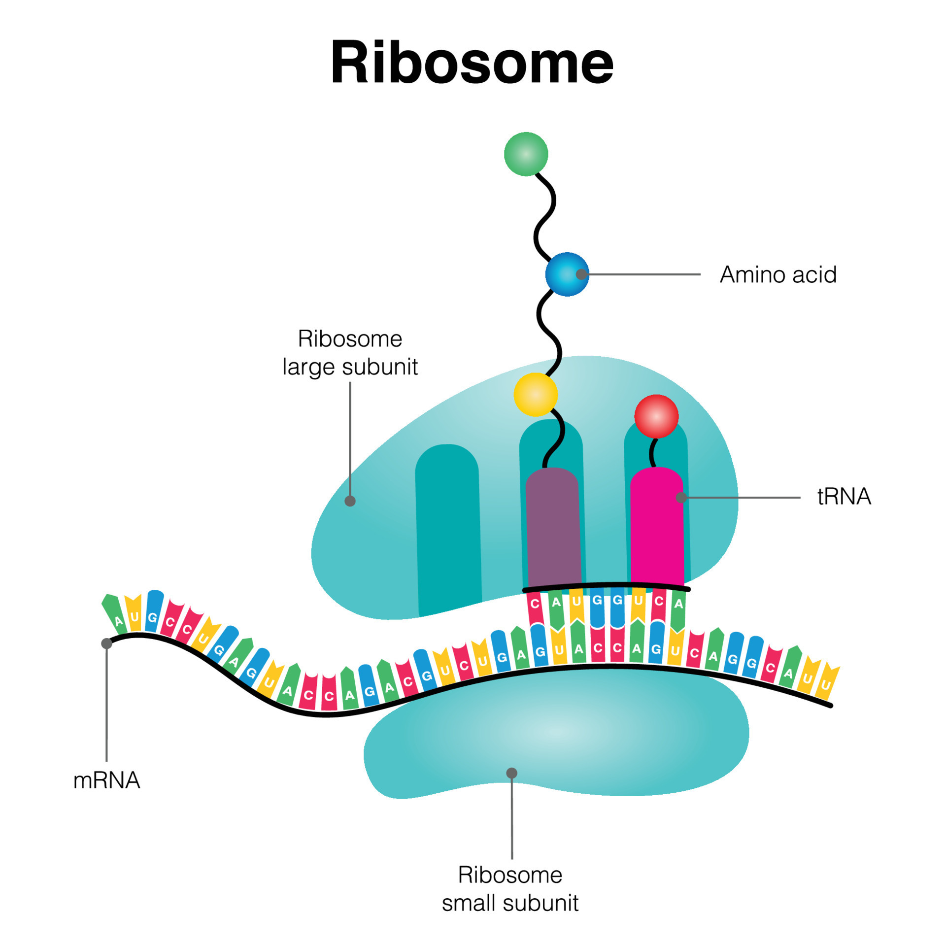
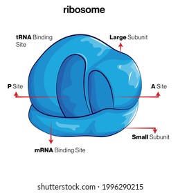
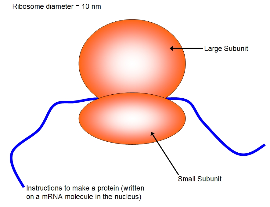
41 ribosome diagram with labels
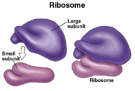
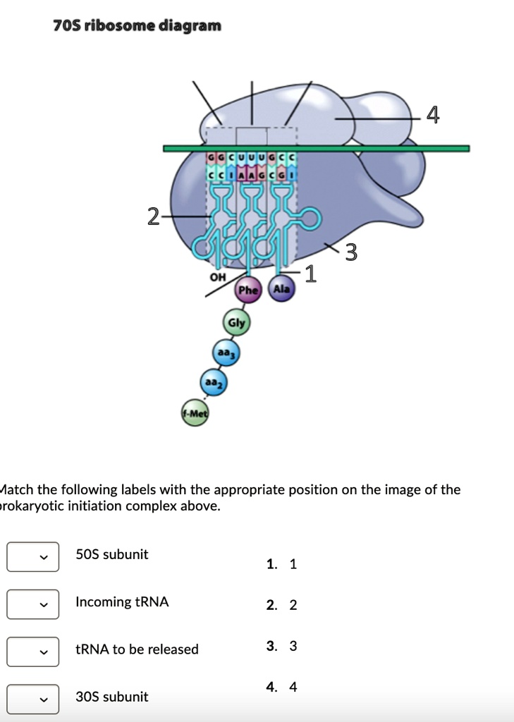
Ribosomes: Structure, Composition, and Assembly (With Diagram) Ribosomes in the cytoplasm of eukaryotic cells have a sedimentation coefficient of about 80 S (MW about 4.5 x 10 6) and are composed of 40 S and 60 S subunits. In prokaryotic cells, ribosomes are typically about 70 S (MW about 2.7 x 10 6) and are formed from 30 S and 50 S subunits. BIONIC: biological network integration using convolutions | Nature … Oct 03, 2022 · Biological networks constructed from varied data can be used to map cellular function, but each data type has limitations. Network integration promises to address these limitations by combining ...
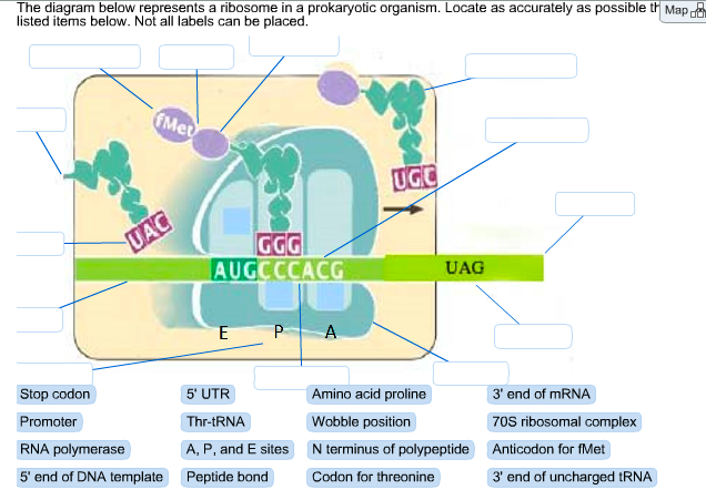
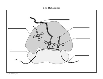
The diagram at right represents a ribosome bound to The diagram at right represents a ribosome bound to mRNA a On the diagram label from LIFESCIENC 7A at University of California, Los Angeles. Study Resources. Main Menu; by School; by Literature Title; ... The diagram at right represents a ribosome bound to mRNA a On the diagram label.

Ribosome diagram with labels
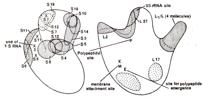
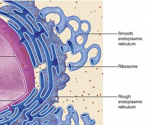
Structure of Subunits of Ribosomes (With Diagram) | Genetics Ribosomes are ribonucleoprotein particles present in all types of cells. They were first observed in EM by Claude in the cytoplasm of cells, and later on the surface of endoplasmic reticulum by Porter and Palade. Ribosomes occur in 3 sizes: 70S in bacteria and chloroplasts, 60S in mitochondria, 80S in cytoplasm of eukaryotes. Ribosome Diagrams A ribosome is a complex of RNA and protein and is, therefore, known as a ribonucleoprotein. It is composed of two subunits - smaller and larger. The smaller. A ribosome functions as a micro-machine for making proteins. Ribosomes are composed of special For an overview diagram of protein production click here. Ribosomes- Definition, Structure, Functions and Diagram - Microbe Notes Ribosomes Definition The ribosome word is derived - 'ribo' from ribonucleic acid and 'somes' from the Greek word 'soma' which means 'body'. Ribosomes are tiny spheroidal dense particles (of 150 to 200 A0 diameters) that are primarily found in most prokaryotic and eukaryotic. They are sites of protein synthesis.
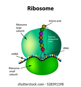
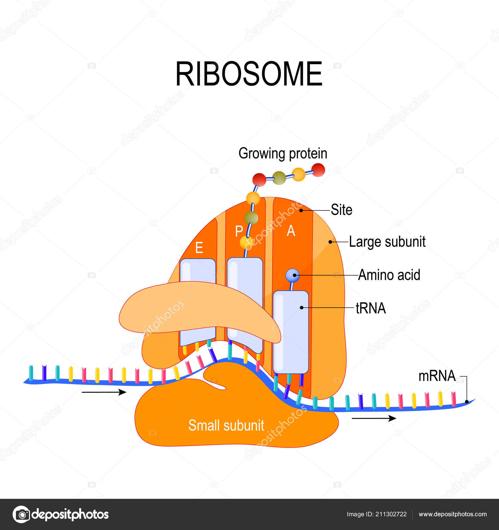
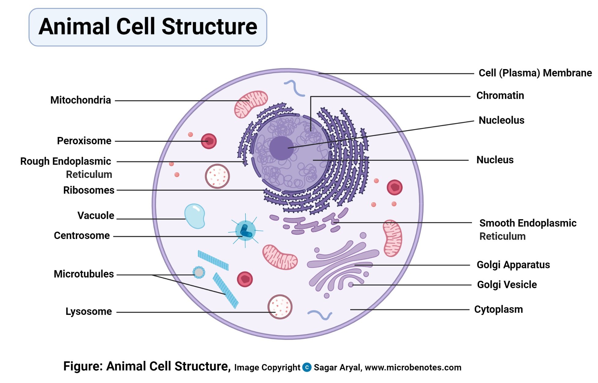
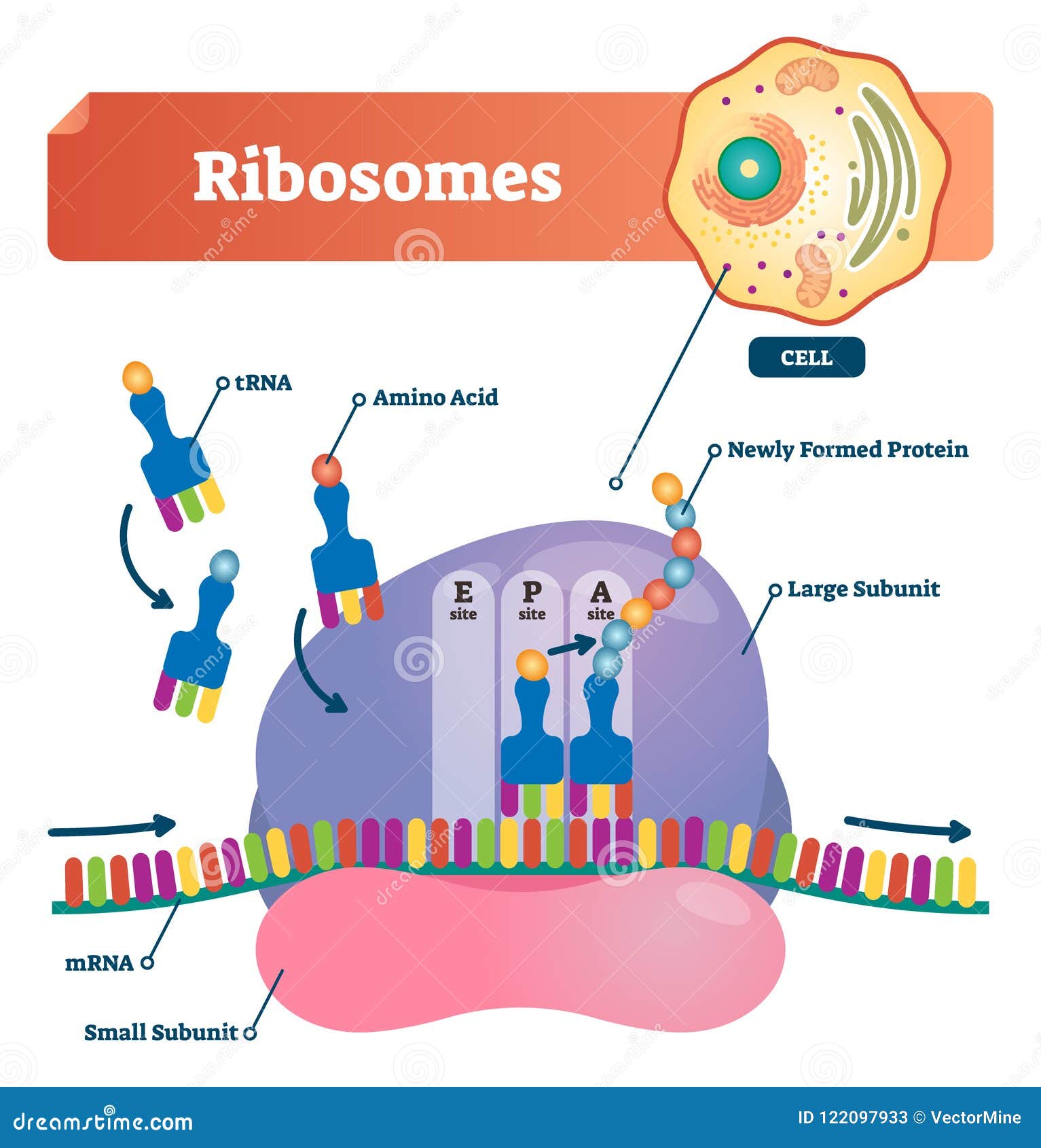
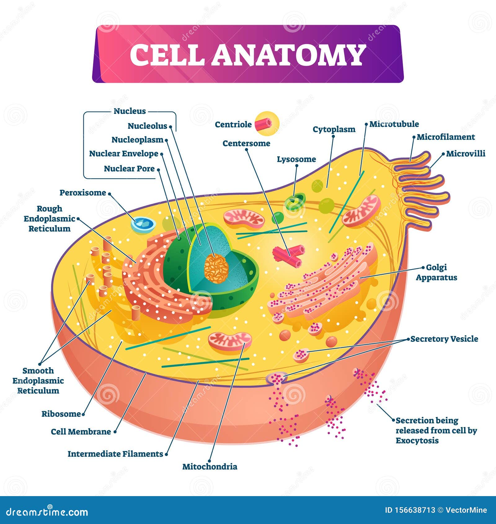
Ribosome diagram with labels. Animal Cells: Labelled Diagram, Definitions, and Structure - Research Tweet Animal Cells Organelles and Functions. A double layer that supports and protects the cell. Allows materials in and out. The control center of the cell. Nucleus contains majority of cell's the DNA. Popularly known as the "Powerhouse". Breaks down food to produce energy in the form of ATP. Molecular Modeling Database (MMDB) Help Document Source Database. The Molecular Modeling DataBase (MMDB) is a database of experimentally determined three-dimensional biomolecular structures, and is also referred to as the Entrez Structure database. It is a subset of three-dimensional structures obtained from the RCSB Protein Data Bank (PDB), excluding theoretical models.The data processing procedure at NCBI results … Ribosome - protein factory - definition, function, structure and biology The protein translation by a ribosome consists of three stages: (1) Initiation, (2) Elongation, and (3) Termination. Initiation - the ribosome assembles around the target mRNA. A small ribosome subunit links onto the "start-end" of an mRNA strand. "Initiator tRNA" also enters the small subunit and binds to the start codon (most commonly, AUG). Solved In the following diagram of a ribosome, assign the - Chegg Question: In the following diagram of a ribosome, assign the correct labels. ... In the following diagram of a ribosome, assign the correct labels. Show transcribed image text Expert Answer. Who are the experts? Experts are tested by Chegg as specialists in their subject area. We review their content and use your feedback to keep the quality high.
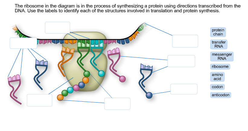
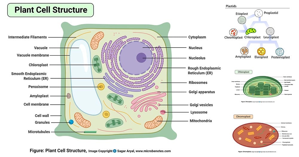
Ribosome - Wikipedia During 1977, Czernilofsky published research that used affinity labeling to identify tRNA-binding sites on rat liver ribosomes. Several proteins, including L32/33, L36, L21, L23, L28/29 and L13 were implicated as being at or near the peptidyl transferase center. [27] Plastoribosomes and mitoribosomes [ edit] Labeled Plant Cell With Diagrams | Science Trends The ribosomes are created in the nucleolus of the cell. Ribosomes are made out of two smaller subunits, a large ribosomes subunit and a small ribosomal subunits. The transfer RNA or tRNA encodes the correct series of genetic instructions into the mRNA or messenger RNA, which is what ensures that the right proteins are created. DNA - Wikipedia Deoxyribonucleic acid (/ d iː ˈ ɒ k s ɪ ˌ r aɪ b oʊ nj uː ˌ k l iː ɪ k,-ˌ k l eɪ-/ (); DNA) is a polymer composed of two polynucleotide chains that coil around each other to form a double helix carrying genetic instructions for the development, functioning, growth and reproduction of all known organisms and many viruses.DNA and ribonucleic acid (RNA) are nucleic acids. Solved The ribosome in the diagram is in the process of | Chegg.com The ribosome in the diagram is in the process of synthesizing a protein using directions transcribed from the DNA. Use the labels to identify each of the structures involved in translation and protein synthesis. Question: The ribosome in the diagram is in the process of synthesizing a protein using directions transcribed from the DNA.
Active Ribosome Profiling with RiboLace - PubMed Ribosome profiling, or Ribo-seq, is based on large-scale sequencing of RNA fragments protected from nuclease digestion by ribosomes. Thanks to its unique ability to provide positional information about ribosomes flowing along transcripts, this method can be used to shed light on mechanistic aspects … Structure of Ribosome - Biology Wise Diameter of Ribosome is 20nm. Their composition can be divided into two parts - 2/3 part of r-RNA (ribosomal RNA) and 1/3 part RNP (Ribosomal protein or Ribonuclep protein). Polypeptide chain is fabricated by translating mRNA (messenger RNA) with the aid amino acids that tRNA (transfer RNA) delivers. Biology 1005 - Chapter 6 DNA: The Molecule of Life - Quizlet Study with Quizlet and memorize flashcards containing terms like Can you complete this paragraph about the cellular processes involved in protein production?, The cellular processes that results in the production of protein begin in the _____ , where the DNA resides. There, the process of _____ creates a molecule of RNA from a molecule of DNA. The enzyme that … Transcription and Translation - YouTube You can learn more about transcription and translation in this course I taught with Udacity: ...
Role of Ribosomes in Protein Synthesis (With Diagram) - Biology Discussion The mRNA binds to the 30S subunit of ribosome to form initiation complex. The main role of ribosome is its ability to catalyse the formation of peptide bonds between amino acids, so that the amino acids are incorporated into proteins. Ribosomes are dense granules without covering membranes. They were first observed by Palade.
Ribosome and protein synthesis, diagram - Stock Image - C029/3020 Messenger ribonucleic acid (mRNA, blue with coloured nucleotides) is read by a ribosome (pink). The molecules of transfer RNA (tRNA, key-shaped) each bring an amino acid (orange dot) to bind to the ribosome's protein synthesis site. The amino acid added is determined by the three-nucleotide coding (codon) on the mRNA, which matches up with the ...
Mastering Quiz: Chapter 7A Microbial Genetics Flashcards | Quizlet Those segments of the RNA strand that do not actually code for the protein are removed. c. mRNA binds to a ribosome in the cytoplasm. d. A molecule of RNA is formed based on the sequence of nucleotides in DNA. e. ... Drag the correct labels under the diagrams to identify the events of RNA processing. Drag the labels onto the diagram to identify ...
Structure of Ribosome (With Diagram) - Biology Discussion A bacterial ribosome is about 250 nm in diameter and consists of two subunits, one large and one small. Both subunits consist of one or more molecules of rRNA and an array of ribosomal proteins. ADVERTISEMENTS: Association of two subunits is called mono-some. The structure of prokaryotic ribosome is given in the figure 8.2 B.
What is the path a secretory protein follows from synthesis to ... Jan 31, 2011 · Protein SynthesisEndoplasmic Reticulum-->cis Golgi cisternae --> medial Golgi cisternae --> trans Golgi Cisternae --> Plasma membraneExtra Cellular SpaceAs they are being synthesized, secretory ...
Ribosomes- Definition, Structure, Functions and Diagram - Microbe Notes Ribosomes Definition The ribosome word is derived - 'ribo' from ribonucleic acid and 'somes' from the Greek word 'soma' which means 'body'. Ribosomes are tiny spheroidal dense particles (of 150 to 200 A0 diameters) that are primarily found in most prokaryotic and eukaryotic. They are sites of protein synthesis.
Ribosome Diagrams A ribosome is a complex of RNA and protein and is, therefore, known as a ribonucleoprotein. It is composed of two subunits - smaller and larger. The smaller. A ribosome functions as a micro-machine for making proteins. Ribosomes are composed of special For an overview diagram of protein production click here.
Structure of Subunits of Ribosomes (With Diagram) | Genetics Ribosomes are ribonucleoprotein particles present in all types of cells. They were first observed in EM by Claude in the cytoplasm of cells, and later on the surface of endoplasmic reticulum by Porter and Palade. Ribosomes occur in 3 sizes: 70S in bacteria and chloroplasts, 60S in mitochondria, 80S in cytoplasm of eukaryotes.
















Post a Comment for "41 ribosome diagram with labels"