39 fluorescent labels and light microscopy
› applications › fretBasics of FRET Microscopy | Nikon’s MicroscopyU The first fluorescent protein biosensor was a calcium indicator named cameleon, constructed by sandwiching the protein calmodulin and the calcium calmodulin-binding domain of myosin light chain kinase (M13 domain) between enhanced blue and green fluorescent proteins (EBFP and EGFP). In the presence of increasing levels of intracellular calcium ... proscitech.com.auProSciTech Laboratory supplies and Lab equipment for Histology, Pathology, Light Microscopy, Electron Microscopy and specialist researchers.
en.wikipedia.org › wiki › Super-resolution_microscopySuper-resolution microscopy - Wikipedia Integrated correlative light and electron microscopy. Combining a super-resolution microscope with an electron microscope enables the visualization of contextual information, with the labelling provided by fluorescence markers. This overcomes the problem of the black backdrop that the researcher is left with when using only a light microscope.
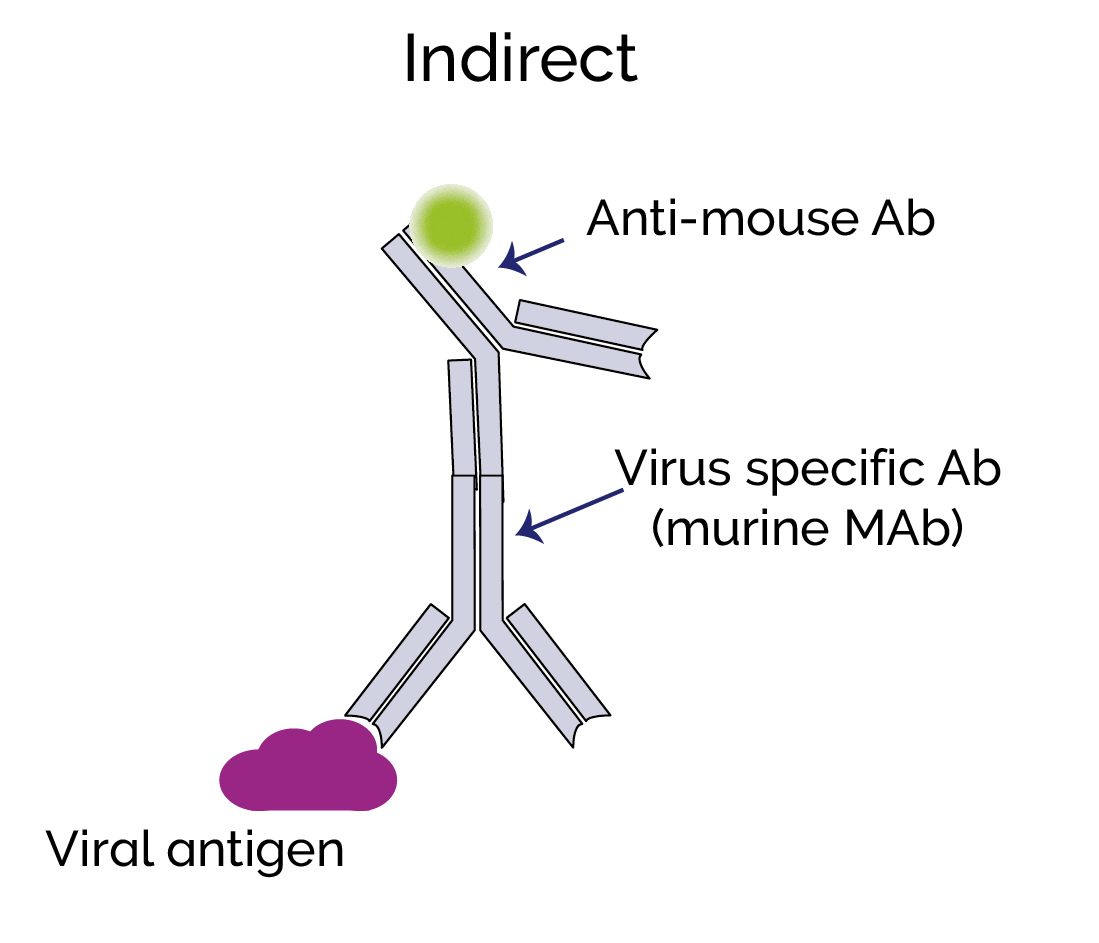
Fluorescent labels and light microscopy
en.wikipedia.org › wiki › Förster_resonance_energyFörster resonance energy transfer - Wikipedia In fluorescence microscopy, fluorescence confocal laser scanning microscopy, as well as in molecular biology, FRET is a useful tool to quantify molecular dynamics in biophysics and biochemistry, such as protein-protein interactions, protein–DNA interactions, and protein conformational changes. For monitoring the complex formation between two ... en.wikipedia.org › wiki › FluorescenceFluorescence - Wikipedia A perceptible example of fluorescence occurs when the absorbed radiation is in the ultraviolet region of the electromagnetic spectrum (invisible to the human eye), while the emitted light is in the visible region; this gives the fluorescent substance a distinct color that can only be seen when the substance has been exposed to UV light ... › articles › s41592/020/01005-2Click-ExM enables expansion microscopy for all biomolecules Dec 07, 2020 · Expansion microscopy (ExM) allows super-resolution imaging on conventional fluorescence microscopes, but has been limited to proteins and nucleic acids. ... Using 18 clickable labels, we ...
Fluorescent labels and light microscopy. › articles › s41467/021/20908-yGeminate labels programmed by two-tone microdroplets ... - Nature Jan 29, 2021 · Guo and Li et al. recently have reported an interesting dual-mode CLC system induced by light-driven fluorescent chiral switches, in which the reflection wavelength and fluorescence intensity were ... › articles › s41592/020/01005-2Click-ExM enables expansion microscopy for all biomolecules Dec 07, 2020 · Expansion microscopy (ExM) allows super-resolution imaging on conventional fluorescence microscopes, but has been limited to proteins and nucleic acids. ... Using 18 clickable labels, we ... en.wikipedia.org › wiki › FluorescenceFluorescence - Wikipedia A perceptible example of fluorescence occurs when the absorbed radiation is in the ultraviolet region of the electromagnetic spectrum (invisible to the human eye), while the emitted light is in the visible region; this gives the fluorescent substance a distinct color that can only be seen when the substance has been exposed to UV light ... en.wikipedia.org › wiki › Förster_resonance_energyFörster resonance energy transfer - Wikipedia In fluorescence microscopy, fluorescence confocal laser scanning microscopy, as well as in molecular biology, FRET is a useful tool to quantify molecular dynamics in biophysics and biochemistry, such as protein-protein interactions, protein–DNA interactions, and protein conformational changes. For monitoring the complex formation between two ...











Post a Comment for "39 fluorescent labels and light microscopy"