38 spinal cord model with labels
Spinal cord transverse section coverings label 3D model Aug 7, 2020 — Spinal cord transverse section coverings label 3D model, available formats , ready for 3D animation and other 3D projects | CGTrader.com. PDF Anatomy & Physiology - TMCC Somso Model KS 4 Block model showing the skin with hair in different planes of section. I. Epidermis II. Corium (Dermis) III. Subcutis (Hypodermis) 1. External Horny Layer (Stratum corneum) 1a. Clear Layer (Stratum lucidum) -(KS 3 only) 2. Internal Hornless Germinative Zone (Stratum germinativum) 2a. Granular Layer (Stratum granulosum) 2b.
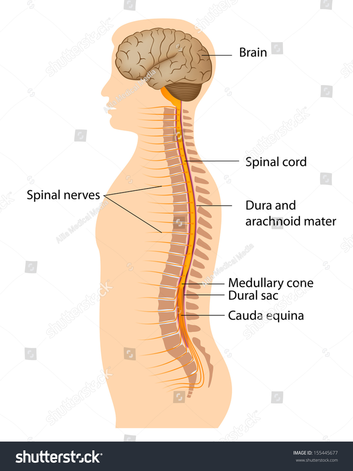
Spinal cord anatomy - Physiopedia The spinal cord is part of the central nervous system and consists of a tightly packed column of nerve tissue that extends downwards from the brainstem through the central column of the spine. It is a relatively small bundle of tissue (weighing 35g and just about 1cm in diameter) but is crucial in facilitating our daily activities.. The spinal cord carries nerve signals from the brain to other ...
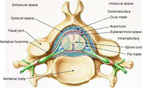
Spinal cord model with labels
Solved Identify the following structures on the spinal cord - Chegg Identify the following structures on the spinal cord model & label the images below. Match the structures with the correct location using lecture notes Show transcribed image text Expert Answer 1.Duramater of spinal cordA 2. Arachnoid mater of spinal cord 3. Piamater of spinal cord 4. 5. 6. White matter 7. Dorsal horn 8. Lateral horn 9. Spinal cord: Anatomy, structure, tracts and function | Kenhub The spinal cord is made of gray and white matter just like other parts of the CNS. It shows four surfaces: anterior, posterior, and two lateral. They feature fissures (anterior) and sulci (anterolateral, posterolateral, and posterior). The gray matter is the butterfly-shaped central part of the spinal cord and is comprised of neuronal cell bodies. Diffusion MRI - Wikipedia Diffusion-weighted magnetic resonance imaging (DWI or DW-MRI) is the use of specific MRI sequences as well as software that generates images from the resulting data that uses the diffusion of water molecules to generate contrast in MR images.
Spinal cord model with labels. Learn the spinal cord with diagrams and quizzes | Kenhub Here you'll simply fill in the blanks with the name of the structure corresponding to the diagram label. You can also download the spinal cord diagram labeled if you'd like to scribble and make notes before attempting the labeling activity. DOWNLOAD PDF WORSHEET (BLANK) DOWNLOAD PDF WORKSHEET (LABELED) Accelerate your learning with quizzes Spine Anatomy, Diagram & Pictures | Body Maps - Healthline To facilitate this process, the spinal cord is divided into two kinds of pathways called tracts. Ascending tracts carry sensory input from the body to the brain, and descending tracts carry... Nervous System Models - Labeled Brain and Spinal Cord Nervous System Models - Labeled Brain and Spinal Cord Anatomy Bones, Gross Anatomy, Brain. Melissa Ritts. 6 followers. More information. Anatomy Bones. Amazon.com: spinal cord model Axis Scientific Spine Model, 34" Life Size Spinal Cord Model with Vertebrae, Nerves, Arteries, Lumbar Column, and Male Pelvis, Includes Stand, Detailed Product Manual and Worry Free 3 Year Warranty 292 $7699 $109.99 Get it as soon as Thu, Oct 6 FREE Shipping by Amazon Small Business More Buying Choices $71.42 (4 used & new offers)
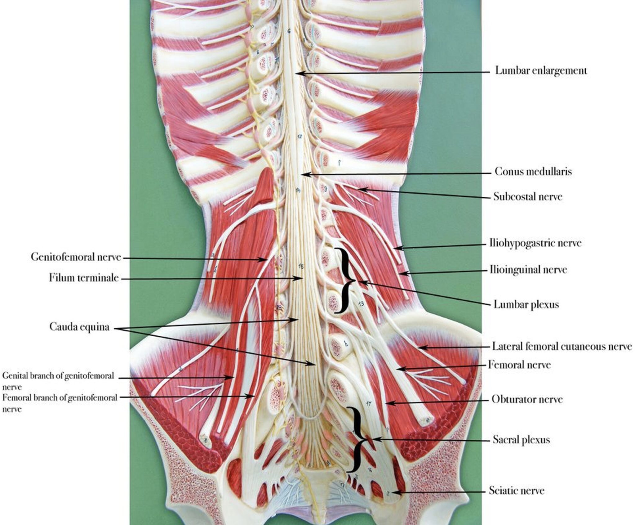
Solved 1. Label this picture of the model: Spinal cord and | Chegg.com 1. Label this picture of the model: Spinal cord and spinal column wall mount • Label: use the free drawing tool/brackets and arrows to Identify the following: • conus medullaris • cauda equina . filum terminale CU ; Question: 1. Label this picture of the model: Spinal cord and spinal column wall mount • Label: use the free drawing tool ... Human anatomy spine labelled royalty-free images Human anatomy spine labelled royalty-free images. 2,836 human anatomy spine labelled stock photos, vectors, and illustrations are available royalty-free. SPINAL CORD MODEL Flashcards | Quizlet Objectives for Spinal Cord (fifth cervic…. 210: Chapter 11 Blended Skills and Critical Thinki…. 5th - Social Studies Review - ch7 notes and questi…. Answered: Can you help me the name of all the… | bartleby Can you help me the name of all the number of the label spinal cord system model? Question. thumb_up 100%. Can you help me the name of all the number of the label spinal cord system model? Transcribed Image Text: 16 19 22 9. 11 Expert Solution. Want to see the full answer? Check out a sample Q&A here.
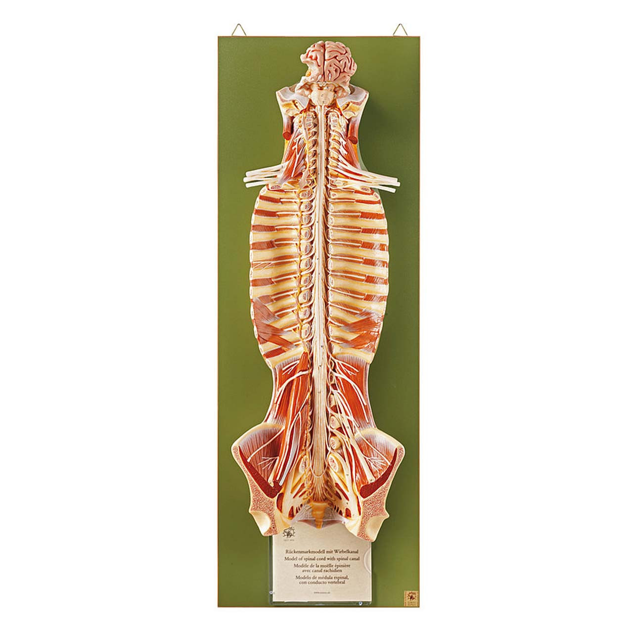
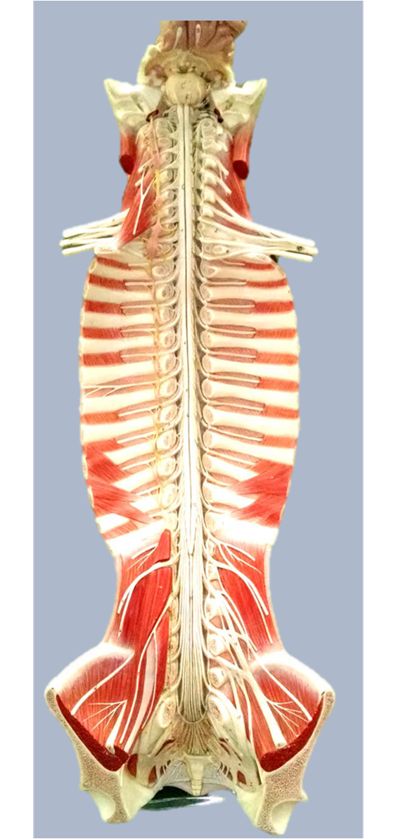
Labeled Human Torso Model Diagram : Biology 2404 A&P Basics ... Labeled human torso models feature clear views of the vertebrae, spinal cord, spinal nerves, vertebral arteries, lungs, stomach, liver, intestinal track, kidneys human anatomical models play an important part in the patient education process. Limited time sale easy return. Ear head torso back upper arm lower arm hand upper leg lower leg foot tail. 21,074 Spinal cord Images, Stock Photos & Vectors | Shutterstock Find Spinal cord stock images in HD and millions of other royalty-free stock photos, illustrations and vectors in the Shutterstock collection. Thousands of new, high-quality pictures added every day. Lab Manual - Deep Back & Spinal Cord - Texas Tech University Health ... Consider the vertebral column as a whole and the structures which unite it: supraspinous ligament, ligamentum flavum, posterior longitudinal ligament, anterior longitudinal ligament (will be seen later), intervertebral disc. Identify intervertebral foramina, superior and inferior vertebral notches, and anterior and posterior sacral foramina. 2. Spinal Cord in the Spinal Canal (BS 31) · Anatomy models - SOMSO® Spinal Cord in the Spinal Canal. Seen from the ventral side, natural size, in SOMSO-PLAST®. The model shows the brain stem and the spinal cord, as well as the nerve branches, up to the coccygeal plexus. On the left side, the sympathetic trunk with its connections to the central nervous system is shown. In one piece. Mounted on a green board.
Spinal cord - Wikipedia The spinal cord is a long, thin, tubular structure made up of nervous tissue, which extends from the medulla oblongata in the brainstem to the lumbar region of the vertebral column (backbone). The backbone encloses the central canal of the spinal cord, which contains cerebrospinal fluid.
Anti-NeuN antibody [1B7] - Neuronal Marker (ab104224) | Abcam Li Y et al. Transcriptome profiling of long noncoding RNAs and mRNAs in spinal cord of a rat model of paclitaxel-induced peripheral neuropathy identifies potential mechanisms mediating neuroinflammation and pain. J Neuroinflammation 18:48 (2021). PubMed: 33602238; View all Publications for this product
Anatomy of the Spinal Cord (Section 2, Chapter 3) Neuroscience Online ... The spinal cord extends from the foramen magnum where it is continuous with the medulla to the level of the first or second lumbar vertebrae. It is a vital link between the brain and the body, and from the body to the brain. The spinal cord is 40 to 50 cm long and 1 cm to 1.5 cm in diameter. Two consecutive rows of nerve roots emerge on each of ...
Spinal Cord Diagram with Detailed Illustrations and Clear Labels - BYJUS Spinal Cord Diagram The spinal cord is one of the most important structures in the human body. It is the most important structure for any vertebrate. Anatomically, the spinal cord is made up of nervous tissue and is integrated into the spinal column of the backbone. Main Article: Spinal Cord - Anatomy, Structure, Function, and Spinal Cord Nerves
spinal cord anatomy, labeling spinal model Quiz - PurposeGames.com This is an online quiz called spinal cord anatomy, labeling spinal model There is a printable worksheet available for download here so you can take the quiz with pen and paper. Your Skills & Rank Total Points 0 Get started! Today's Rank -- 0 Today 's Points One of us! Game Points 16 You need to get 100% to score the 16 points available Actions
Spinal Cord sections labeled Diagram | Quizlet Start studying Spinal Cord sections labeled. Learn vocabulary, terms, and more with flashcards, games, and other study tools.
Spinal cord transverse section coverings label - Sketchfab Jan 4, 2020 — A blend model of spinal cord along with it covering layers and nerve roots. The ascending and descending tracts of spinal cord transverse ...
Labeled Brain Model Diagram | Science Trends The medial region of the posterior and anterior lobes function to control fine body movements, taking in input from the spinal cord as well as the auditory and visual systems of the brain. The lateral region of the cerebellum is the largest part of the cerebellum in humans. This region gets inputs from the cerebral cortex.
spinal cord anatomy, labeling spinal model - Printable - PurposeGames.com This is a free printable worksheet in PDF format and holds a printable version of the quiz spinal cord anatomy, labeling spinal model. By printing out this quiz and taking it with pen and paper creates for a good variation to only playing it online.
Nervous System Models - Labeled Brain and Spinal Cord - Pinterest Feb 4, 2017 - Nervous System Models - Labeled Brain and Spinal Cord. Feb 4, 2017 - Nervous System Models - Labeled Brain and Spinal Cord. Feb 4, 2017 - Nervous System Models - Labeled Brain and Spinal Cord. Pinterest. Today. Explore. When autocomplete results are available use up and down arrows to review and enter to select. Touch device users ...
Spinal Cord Models - San Diego Mesa College Spinal Cord Models. photo for a larger view of the model. Click on Label for the labeled model. Back to Nervous System. Spinal Cord. (transverse section) Spinal Cord (close up) Spinal Cord. (longitudinal view)
The FASEB Journal - Wiley Online Library Dec 27, 2021 · We are delighted to welcome Dr. Jeannine Botos to FASEB in the newly created role of Senior Managing Editor for The FASEB Journal and FASEB BioAdvances. Jeannine received a B.S. in Biochemistry and a Ph.D. in Veterinary Physiology and Pharmacology from Texas A&M and completed a postdoc at the National Cancer Institute.
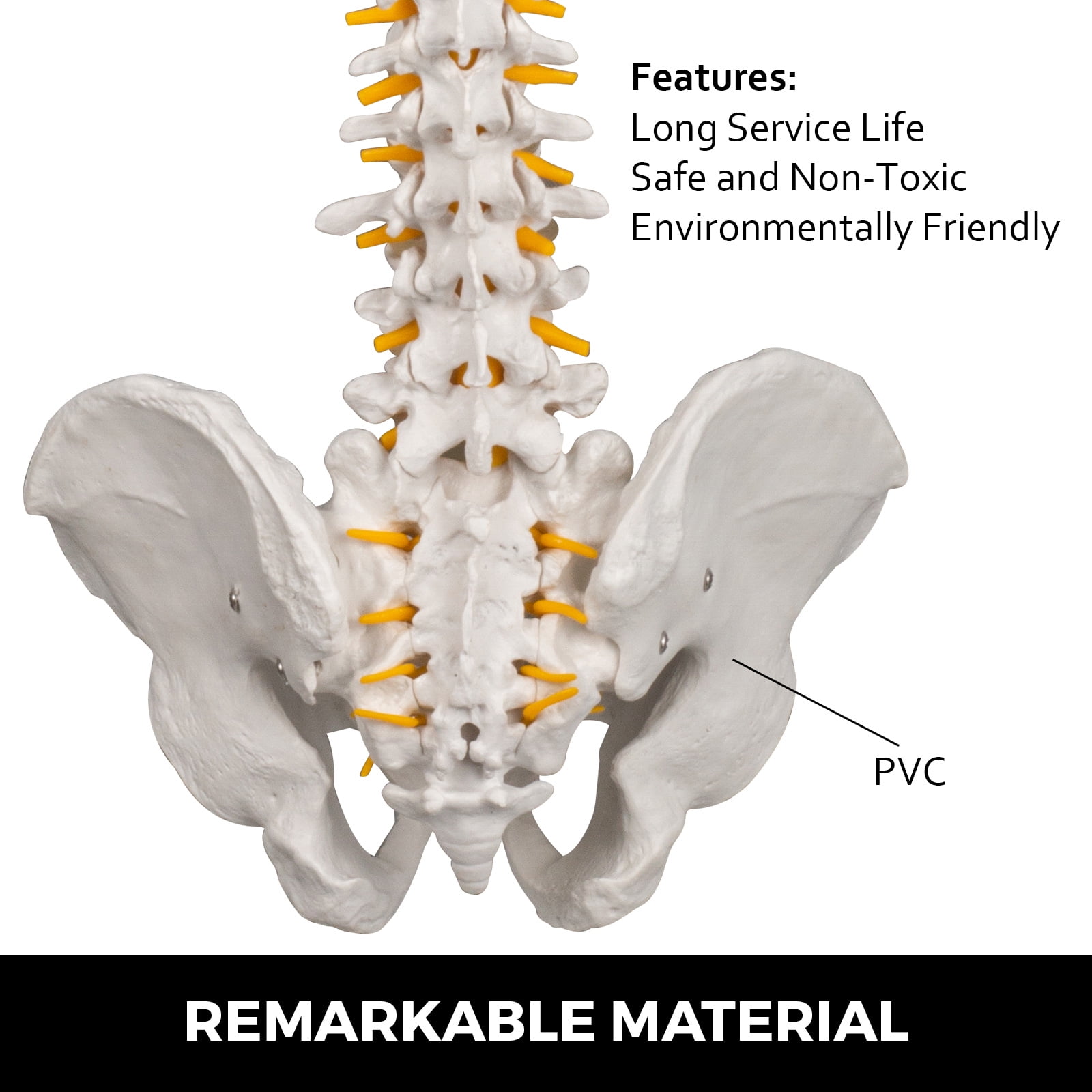
VEVOR White Vertebral Column Model 33inch Total Height Skeleton Spine Model 30inch Spine Length Life Size Spine Model with Spinal Nerves, Skull Base ...
Spinal Cord - Indiana University Bloomington The Cell and Cell Division: Spinal Cord. Spinal Cord . Last updated: 31 July 2005 Comments: Dr. Valerie Dean O'Loughlin, Matthew William Crosby Copyright 2002, The ...
Axis Scientific Spine Model, 34" Life Size Spinal Cord Model with ... Axis Scientific Spine Model, 34" Life Size Spinal Cord Model with Vertebrae, Nerves, Arteries, Lumbar Column, and Male Pelvis, Includes Stand, Detailed Product Manual and Worry Free 3 Year Warranty ... Full Color Spine Model Study Guide . Includes 26 labeled parts! Read more. Read more. Read more. Brief content visible, double tap to read full ...
Spinal Column and Spinal Cord - 3D Model - MSD Manuals Brought to you by Merck & Co, Inc., Rahway, NJ, USA (known as MSD outside the US and Canada)—dedicated to using leading-edge science to save and improve lives ...
Spinal Cord - Anatomy, Structure, Function, & Diagram - BYJUS In adults, the spinal cord is usually 40cm long and 2cm wide. It forms a vital link between the brain and the body. The spinal cord is divided into five different parts. Sacral cord Lumbar cord Thoracic cord Cervical cord Coccygeal Several spinal nerves emerge out of each segment of the spinal cord.
Q-Link Acrylic SRT-3 Pendant (Original Black) - amazon.com Manufacturer direct listing + FREE 2-3 Day Shipping for USA orders ; The Premier Pendant for Protection Against Negative EMF Effects ; Package includes 1 brand new sleek, stylish, durable Q-Link Acrylic SRT-3 Pendant made in USA + 30” comfort cord
Spinal Cord Quiz: Cross-Sectional Anatomy | GetBodySmart Spinal Cord - Cross-Sectional Anatomy. Start Quiz. Want to learn faster? Look no further than these interactive, exam-style anatomy quizzes. Learn anatomy faster and remember everything you learn. Start Now. Related Articles. Parts of the Brain Quiz. Test your knowledge with the parts of the brain and their functions in a fun and interactive ...
PDF Anatomy and Physiology of the Spinal Cord The spinal cord is a bundle of spinal nerves wrapped together. The spinal nerves enter and exit the spinal cord through small spaces between the vertebrae. The blood vessels which carry oxygen to the spinal cord also use these spaces. You have 8 pairs of cervical nerves, 12 thoracic, 5 lumbar and 6 sacral.
Spinal Cord Labeled Pictures, Images and Stock Photos Browse 154 spinal cord labeled stock photos and images available, or start a new search to explore more stock photos and images. Newest results Spinal stenosis vector illustration. Labeled medical scheme with... Spinal stenosis vector illustration. Labeled medical scheme with explanation.
Diffusion MRI - Wikipedia Diffusion-weighted magnetic resonance imaging (DWI or DW-MRI) is the use of specific MRI sequences as well as software that generates images from the resulting data that uses the diffusion of water molecules to generate contrast in MR images.
Spinal cord: Anatomy, structure, tracts and function | Kenhub The spinal cord is made of gray and white matter just like other parts of the CNS. It shows four surfaces: anterior, posterior, and two lateral. They feature fissures (anterior) and sulci (anterolateral, posterolateral, and posterior). The gray matter is the butterfly-shaped central part of the spinal cord and is comprised of neuronal cell bodies.
Solved Identify the following structures on the spinal cord - Chegg Identify the following structures on the spinal cord model & label the images below. Match the structures with the correct location using lecture notes Show transcribed image text Expert Answer 1.Duramater of spinal cordA 2. Arachnoid mater of spinal cord 3. Piamater of spinal cord 4. 5. 6. White matter 7. Dorsal horn 8. Lateral horn 9.


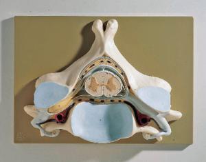
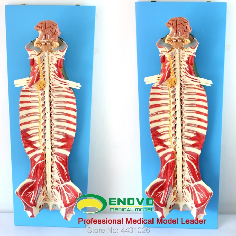





:background_color(FFFFFF):format(jpeg)/images/library/11473/spinal-membranes-and-nerve-roots_english.jpg)




Post a Comment for "38 spinal cord model with labels"